AVR Dragon Programmer / Debugger by Atmel Unboxing

Working with the Arduino is a lot of fun, and it is a great way to get your feet wet in the world of microcontrollers.  But when you decide to start working on more customized projects, you will realize that Atmel makes a lot of different AVR microcontrollers and that you will need a way to program them rather than using a bootloader.

That’s where the AVR Dragon programmer / debugger from Atmel comes into play.  The Dragon is inexpensive at about $49 plus shipping (Atmel store page).  Because it is inexpensive, it does not come with any accessories – or so it seemed.  One person told me that it came with cables.  Another told me that it came with a single 10-pin Socket/Socket IDC cable.  Because I wasn’t sure what might or might not come with the Dragon, I didn’t order any accessories until it arrived.

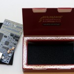
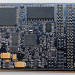
So for other people who may be curious about exactly what is in the box, here is the unboxing of the AVR Dragon that I received on September 10th, 2013.


As you can see, the box contains only the programmer, no box, cables, or power supply.  Next up Outfitting the Dragon AVR Programmer.

Share
Posted in Atmel AVR microcontrollers, Programming | Comments Off on AVR Dragon Programmer / Debugger by Atmel Unboxing

S.1627 — Resident Physician Shortage Reduction Act of 2011

On October 6th, 2011, the New England Journal of Medicine published a Health Policy Report entitled “The Uncertain Future of Medicare and Graduate Medical Education“.  The Report is worth reading, but the take home message is that we will soon have people graduating from medical school who are unable to find a residency position.  A physician who hasn’t completed a residency can’t get a medical license in the United States.

For those of you that aren’t familiar with why a residency position is important, it’s because medical education works like this:

After obtaining a four year Bachelors degree from a University, you attend an additional four years of medical school, typically two years of didactic (lecture) learning, followed by two years of clinical rotations (hospital education).  Upon graduation from medical school, you are awarded the Doctor of Medicine (MD) degree.  At this point, you have the MD after your name, but your practical experience is still limited to the two years of clinical rotations.

Once you have completed your MD degree, you need to make a decision as to which specialty you will pursue.  Different specialties take different types of training.  A family practice physician pursues a different path than does an internal medicine physician, who takes a different path than a physician who specializes in Emergency Medicine.  This specialized training is known as a “residency”, after the days when the training physicians lived at the hospitals where they were training.  Without completing a residency, an MD is unable to become licensed, and therefore can’t treat patients – effectively ending their career as a physician before it ever begins.

Here is the problem:  Training a resident costs a hospital money.  While some people see residents as low-cost labor for the hospital, it still takes time for the senior physicians (“attendings”) to supervise the residents.  Think of it like this:  Imagine that you are making dinner for a group of friends who are coming over for a visit.  You know that it will take you a set amount of time to make dinner and have it ready for when your friends arrive.  Now imagine that your 10 year old nephew is going to make the dinner for you while you supervise his cooking.  Will the free labor of your 10 year old assistant make dinner come together faster or slower?  How do you think dinner will taste?  Now imagine that instead of having a 10 year old assistant to make dinner, you have a freshly minted, MD qualified physician.  Sounds pretty good, right?  Now instead of making dinner, change your scenario to a kidney transplant.  Now imagine that you are the person that is on the operating room table, getting cut open to get your new kidney.

Obviously, it will require more supervision to train a resident than for the attending physician to simply do the job themselves.  In order to encourage hospitals to operate residency programs, the Federal government pays a little bit extra for every procedure (covered by Medicare/Medicaid)  done in a hospital with a residency program.  The funding for residency programs has been frozen for the past decade, so hospitals have no incentive to increase the size of their residency programs.

The need for new physicians is well known.  As the boomers age, those physicians that grew up with them are starting to retire.  The average age of a US physician is over 45 years old, and many are approaching retirement age.  Medical schools have started to increase their class size and several new medical schools have opened in the past few years.

But because residency funding hasn’t been increased in over 10 years, teaching hospitals are not increasing the size of residency programs.  We are approaching a glut of medical school graduates who won’t be able to find a residency program, leaving America with a shortage of trained physicians and medical school graduates unable to practice.

Help may be at hand in the form of Senate Bill S.1627 — Resident Physician Shortage Reduction Act of 2011.  This Bill calls for an increase in the number of residency positions by 15,000 over the next five years – 3,000 new positions per year.  But once again, there is a catch – how will the increase in residency positions be funded?  After its introduction, S.1627 has been read to the Senate twice and was then referred to the Committee on Finance.

Without action on your part, S.1627 could die an unnoticed death in the hands of the Committee on Finance.  Contact your Senator today and ask for them to support the passage of S.1627.  The Committee on Finance is in session right now (October 2011) – your support could be the vote that makes sure S.1627 gets funding.

To contact your Senator, first look up your State at Senate.gov (look in the upper right hand corner of the page).  You will have two Senators listed, and they will probably have a link to their web contact form on the page.  Use their contact form to express your support for S.1627.  Use your own words – form letters don’t count as much as the words of individuals.  You can be as short or verbose as you choose.  A simple “Please support S.1627” is much better than no message at all.  Click on Senate.gov and support S.1627 — Resident Physician Shortage Reduction Act of 2011.

Share
Posted in Biology | Comments Off on S.1627 — Resident Physician Shortage Reduction Act of 2011

How to learn to read EKGs

I’ve had several people ask me about learning to read EKGs.  In my opinion, there are two things that you must master to effectively interpret EKGs.

First, and most obviously, you must understand the conduction system of the heart, the electrical concepts behind obtaining an EKG, and the normal EKG waveform and intervals.  I think of this as the “mechanical” part of reading an EKG, where the knowledge is well defined and is simply a matter of learning.

Second, and not so obviously, you have to be able to analyze a real world EKG by extracting the important information and ignoring the distractions.  An EKG is like a painting by Salvador Dali, it probably contains more information than you need to know.

The Hallucinogenic Toreador (1968–1970) by Salvador Dalí

What is important information?  Things that you need to know for your specific requirements: rate, rhythm, ST segment changes, signs of electrolyte imbalances or drug overdoses.  Things that are not important depend on your situation at the time – if you are working in a busy ER, and a patient comes in with massive ST segment changes, you probably don’t need to worry about the low level noise seen on the EKG (was the patient shivering?, were the lead wires moving?, is there a loose connection on the grounding wire?), you just need to get the patient to the cath lab NOW.

The only good way to learn how to pull out the information you require from the background noise is by reviewing EKGs.  Lots and lots of EKGs.

For the first part of learning EKGs, the mechanical section, I recommend a book by Brenda Beasley, Understanding EKGs: A Practical Approach (3rd Edition).  If you read the reviews of Beasley’s book, one common complaint is that it is too simplistic, and that it doesn’t have enough review strips.  That isn’t actually a bad thing, because this book is one of the best at teaching you the mechanics of an EKG, and we can leave the information extraction experience to a different book.

For the second half of learning EKGs, we turn to a book by Jane Huff, ECG Workout: Exercises in Arrhythmia Interpretation (Huff, ECG Workout).  This is a book of over 600 EKG strips, with all the messiness of the real world.  Here you will find not just important rhythms, but also pens that smear ink, patients that are shivering, and all the other gems found during the practice of medicine.  Some of the reviews mention that the Huff workbook it too advanced, that it is too hard for beginners.  But that’s OK, because we already got the basics from Barbara Huff’s book.

When you are evaluating EKGs, you will need a good set of calipers that won’t slip when you are making measurements.  These Prestige calipers from Amazon will work well for getting started: Prestige EKG Caliper.

Interpreting EKGs can be fun, just like reading a book, where the lines on the page tell a story to those that can read the words.  Let me know of any interesting rhythms that you run across!

Share
Posted in Biology, Medicine | Comments Off on How to learn to read EKGs

Etymology of “Clostridia perfringens”

What do names mean?  Why is a dog named “dog”?  These are questions that have perplexed three year old children for eternity – and vexed their parents.  In the world of microbiology, organisms are usually named for the people who discovered them, or for a characteristic of the organism.  For example, Kingella kingae, a small, Gram-negative coccobacilli that is a common cause of endocarditis is named after the American bacteriologist Elizabeth O. King.  Alternatively, an organism can be named after something that it causes, such as Vibrio cholerae, which causes the diarrheal disease cholera.

Clostridia are a genus of Gram-positive, rod shaped bacteria.  The genus name, Clostridium, is derived from the Greek term kloster, meaning “spindle”, which fits the spindle-like shape of the organism.  Clostridium perfringens is a pathogenic species of Clostridium that causes a wide range of disease in humans – from a limited gastroenteritis to a myonecrosis termed gas gangrene.  It is from gas gangrene that Clostridium perfringens gets its name.  Perfringens is derived from the Latin, per: “through,” and frango: “to break in pieces, shatter”.

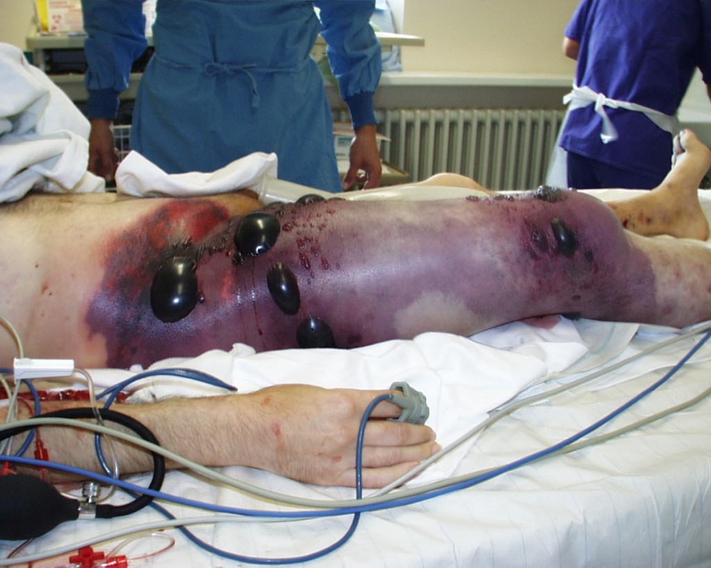
When it occurs, the onset of gas gangrene usually begins within a week of infection with C. perfringens.  C. perfringens encodes at least 12 endotoxins, of which one, Alpha toxin, is a lecithinase.  Lecithin is a general term for a group of lipids (fats) used in the cells of plants and animals.  Alpha toxin specifically cleaves the phospholipid phosphatidylcholine found in the cell membrane of animal cells.  Cleavage of the membrane phosphatidylcholine results in the production of diacylglycerol.  Diacylglycerol activates the protein kinase C second messenger system.  The end result of the second messenger system is a massive increase in inflammation and edema, with devastating and often fatal results.

So now we know how Clostridium perfringens got its name.

The right thigh of a patient suffering from gas gangrene secondary to infection with Clostridium perfringens. Note the massive edema produced by the triggering of the second messenger pathway by Alpha toxin. This patient died 8 hours after admission to the hospital. Image courtesy of: Engelbert Schröpfer, Stephan Rauthe and Thomas Meyer. doi:10.1186/1757-1626-1-252

Share
Posted in Biology | 1 Comment

Books to learn by in my first term at medical school.

The first term of medical school was a challenge.  Biochemistry had changed significantly since I took the course as an undergraduate.  Comparative anatomy was a distant memory of preserved animals and the smell of formaldehyde.

Then, there is my learning style – I am a tactile and visual learner, and I have an absolute requirement to be able to dig deep into the underpinnings of a concept before I can understand the idea fully.  I often find books that start of with “Essentials of …” or “A Review of …” to be watered down versions of the real thing – a problem for me because the text then skips over important sections with no notice to the reader.  Therefore, I tend to like books that cover a topic to a deeper level than required for the courses.  It is important to me that my reference materials not be the limiting factor for my learning.  As an additional factor, I really appreciate good writing and editing.  A well written and edited book is a joy to read.

Here is my list of books that I found to be essential additions for my first term of medical school:

Biochemistry:  I found Lehninger’s “Principles of Biochemistry” to be just what I needed, in-depth, concise, and extraordinarily well written.

Because I am a visual learner, I really got a lot out of The Anatomy Coloring book. When paired with the bright colors of the Staedtler triplus fineliner pens, I was able to learn anatomy and still stay inside the lines. True artists would probably enjoy using colored pencils because the Staedtler pens don’t shade well. But for me, bright primary colors help me remember the layout of the brachial plexus.

Embryology can be a challenge.  It is a highly 3D process that is described using text and 2D images.  I found Langman’s Medical Embryology to be the most helpful for me.  It also comes with a link to a website that gives you some 3D animations, which were nice to help me wrap my head around the process.

I have other recommendations for the second term of medical school that will be coming in another post.

Share
Posted in Biology | Comments Off on Books to learn by in my first term at medical school.

How to get pushed around inside a cell

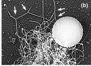
Polystyrene Bead being pushed by Actin

Polystyrene Bead being pushed by F-actin filaments

What do Listeria monocytogenes (a Gram positive rod), Shigella dysenteria (a Gram negative rod), and the Vaccinia virus (a double stranded DNA poxvirus) all have in common?  They all hijack the actin polymerization capability of the host cell in order to become motile.

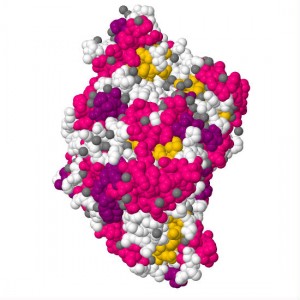
Actin is a globular, somewhat “U” shaped molecule that is used for many different functions in the cell (a moonlighting molecule).  When actin exists as a monomer, it is known as G-actin, short for Globular actin.  The aspect of actin that is exploited by the pathogens is the ability of actin to form long, branched, fibers.

Globular molecule of G-actin

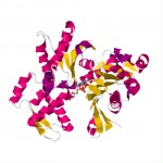
Globular molecule of G-actin. PDB 3HBT.

When G-actin is polymerized into a fiber, it is known as F-actin, short for Filamentous actin.  F-actin is polarized, with one end barbed and the other end pointed.  The F-actin fiber grows from the barbed end and tends to dissociate at the pointed end.  Growth of the fiber is accomplished by having an ATP bound G-actin bind to the barbed end.  About two seconds after binding to the fiber, the ATP is hydrolyzed to ADP, and about six minutes after binding the ADP-G-actin complex dissociates from the F-actin filament.  The rate of fiber extension or dissolution can be changed by cellular enzymes, thereby giving the cell control of the fiber growth.

F-actin filament

F-actin filament made up of G-actin.

Fibers can also branch by using Actin Related Proteins (ARP) 2 and 3.  ARP2/3 binds to a fiber and starts the growth of a new fiber.  This branching creates a tangle of fibers that has a large amount of drag in the intracellular environment.  By extending F-actin from the tangle of fibers a force can be generated that can act upon an object.  Using an enzyme that catalyzes the extension reaction, a force can be generated in one direction.  In this way, the growing tangle of F-actin pushes the enzyme coated object forward.  Mammalian cells use the enzyme profilin placed upon a cell membrane to create a pseudopod structure.

It is important to note that the F-actin fibers themselves do not move, but simply continue to elongate and thereby pushes the catalytic object.  Because the F-actin dissociates after a while, the tangle spontaneously dissolves.  The released G-actin molecules can be reactivated by ATP and can then move back to the head of the fiber.

Close-up of a polystyrene bead coated on one side with ActA

Polystyrene bead with ActA coating on 37.5% of surface area showing F-actin filament formation associated with ActA

Lysteria monocytogenes uses a different enzyme, Actin assembly-inducing protein (ActA), to induce the extension of F-actin filaments.  By expressing ActA at only one end, the rod shaped L. monocytogenes is able to be pushed forward.  As seen in the attached images, the pushing action can be replicated by coating a polystyrene bead with ActA on one side and putting the coated bead in a cell free solution that contains G-actin and ATP.

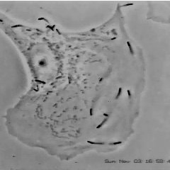
The pushing action induced by Listeria monocytogenes is shown in this excellent video from the lab of Dr. Julie Theriot by Julie Theriot & Dan Portnoy.

Video of Listeria monocytogenes moving in PtK2 cells. --Julie Theriot & Dan Portnoy

For a more complete review of this topic, I heartily suggest Dr. Julie Theriot’s lecture series at iBioSeminars, especially Part 1: Cell Motility.  Her 44 minute lecture will give you a relatively complete understanding of how cells move – an understanding that is truly critical to understanding the world around us.

Here is a YouTube video of Vaccina virus moving inside a cell.  The Vaccinia virus is labelled with green fluorescence and the actin filaments are red.  Unlike L. monocytogenes, Vaccinia does not have a strongly polarized coating of F-actin polymerizing enzyme.

Another excellent reference is the work of the Liu Research Group, headed up by Dr. Andrea Liu.

Clearly, this has been a cursory summary of this important topic.  My thanks go out to all of those researchers who have spent their precious time placing bricks on the tower of knowledge.

Share
Posted in Biology, Medicine | Comments Off on How to get pushed around inside a cell

Pacemaker implant in Grenada

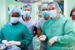
Pacemaker Implant Team at the Grenada General Hospital

Pacemaker Implant Team at the Grenada General Hospital. Photo credit: Josh Yetman

On May 20th, 2011, Dr. Jason S. Finkelstein implanted a heart patient with a dual chamber pacemaker at the Grenada General Hospital.  I was honored to be asked to assist with the implant, based upon my previous experience with cardiac rhythm management devices.

Pacemaker implants are rare in Grenada, with only about 25 devices implanted over the past 10 years.  Dr. Mark Lanzieri pioneered the implant program and continues to implant devices during his visits to Grenada.

Dr. Johansen Sylvester is the driving force behind St George’s University’s Visiting Cardiology Program, which brings cardiac care to the citizens of Grenada.

The patient was in third-degree atrioventricular heart block, with a ventricular rate of about 35 bpm.  Once the pacemaker was implanted, the device began to track the patient’s P wave and pace the ventricle in response to the patients intrinsic atrial rate.

The device was a St. Jude Medical Identity® ADx dual chamber pacemaker, paired with the Tendril® SDX series of lead wires.

Share
Posted in Biology, Medicine | Comments Off on Pacemaker implant in Grenada

Parkinson’s disease: phase II gene therapy clinical trials show promise

Parkinsonism is a syndrome which is characterized by resting tremor, bradykinesia, rigidity, and postural instability.  While there are many different causes of parkinsonism, the most common cause is Parkinson’s disease (PD).  Parkinson’s disease is a neurodegenerative disease that occurs when cells in the substantia nigra (pars compacta) section of the midbrain that normally produce and deliver dopamine to the neostriatum begin to die.  When somewhere around 70% of the dopamine producing cells in the substantia nigra have died, the symptoms of parkinsonism start to appear.

Most pharmacological treatments for PD have focused on increasing the amount of dopamine that is produced in the brain (levodopa), or by stimulating the receptors for dopamine directly using a drug that mimics dopamine (ropinirole, pramipexole).  While these approaches can help with the symptoms of PD, the disease still progresses over the course of 10 to 15 years to the point of virtual immobility.

The hope and promise of gene therapy has been the idea of delivering a gene to a cell, having that cell express the protein encoded by the gene, and having the expressed protein doing something that will accomplish the goals of the people who delivered the gene to the cell.

The good people at Neurologix, Inc. (OTC Bulletin Board: NRGX) seem to have done just that with respect to a gene that can help with PD.  According to a paper published in The Lancet Neurology, AAV2-GAD gene therapy for advanced Parkinson’s disease: a double-blind, sham-surgery controlled, randomised trial, patients who received the therapy showed a significantly improved baseline over patients who received the sham surgery.  Neurologix used an adeno-associated virus (AAV) as the delivery system along with a gene encoding glutamic acid decarboxylase (GAD) as the payload.  The therapy was delivered bilaterally to the subthalamic nuclei using a catheter mechanism.

The wonderful thing about this therapy is the hope that the effects will be ongoing, without the progressive decline in efficacy seen in current therapies.  Although none of the patients were reported to be back to a baseline level of operation, the ability to move slowly is much better than not being able to move at all.

The success of the Phase II clinical trial opens the door to a Phase III clinical trial with many more patients.  I’m sure that many of us are looking forward to the results of the Phase III trial.

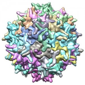
Image of an Adeno-Associated Virus

Image of a Glutamic Acid Decarboxylase molecule

Share
Posted in Biology, Medicine | Comments Off on Parkinson’s disease: phase II gene therapy clinical trials show promise

beta-carboline: Do we learn better when we are anxious?

I’m currently studying the sedative-hypnotic group of drugs.  There is a great deal of chemical variation within the group, so a drug is classified as a sedative-hypnotic based upon it’s clinical effects, and not on it’s chemical makup.

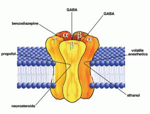
One class of drugs within the sedative-hypnotic group is the benzodiazepines.  The benzos work by binding to a receptor site between an alpha subunit and a gamma subunit on a GABAA receptor chloride channel, where they potentiate the action of GABA by increasing the frequency of opening of the GABAA chloride channel.  The GABAA channel is constitutively active, which means that it opens and closes on its own at a regular rate.  When different chemicals bind to receptor sites on the GABAA channel, they can change the way the channel opens or closes.

Pentameric GABA Receptor <br>Image credit: US National Institutes of Health

Pentameric GABA Receptor Image credit: US National Institutes of Health

The benzodiazepines are agonists, that is, they help open the GABAA chloride channel.  As mentioned above, they increase the frequency at which the GABAA chloride channel opens.  A few common benzodiazepines are diazepam, clonazepam, and lorazepam.

The barbiturates are also agonists.  Unlike the benzodiazepines, they don’t change the frequency of at which the GABAA channel opens.  Instead, they increase the amount of time that the channel stays open once it is activated.  Common barbiturates are phenobarbital, pentobarbital, and thiopental.

GABA agonists all have similar actions: sedation, anticonvulsant effects, muscle relaxation, and anesthesia.

Another drug, flumazenil, is an competitive antagonist of the GABAA receptor at the benzodiazepine binding site and is used as an antidote to benzodiazepines and the non-benzodiazepine benzodiazepine-receptor agonists (NBBRAs).

Now we come to the drugs which are the actual focus of this post, the beta-carbolines.  The beta-carbolines are inverse agonists of the GABAA receptor function.  They block the normal, constitutive, action of the GABA chloride channel.  So they decrease the amount of chloride that enters the cell, reducing the polarization of the neuron, and increasing the chance that the neuron will fire.  As we would expect from a chemical that has the opposite effect of an agonist, beta-carbolines can cause anxiety and seizures.

But the beta-carbolines also seem to have another effect: increasing learning performance and memory.  According to this article from 1986, Benzodiazepine impairs and beta-carboline enhances performance in learning and memory tasks, beta-carbolines help us learn and remember.

While we may not have access to the USP version of the chemical used in the article above, there is another readily available source of beta-carbolines: coffee.

beta-carboline

Share
Posted in Biology, Medicine | Comments Off on beta-carboline: Do we learn better when we are anxious?

Nesidioblastosis

Acquired hyperinsulinemia, usually secondary to gastrointestinal bypass surgery.


Share
Posted in Medicine | Comments Off on Nesidioblastosis